
Parametric oscillators
A core component of many of the group's experiments is the parametric oscillator.

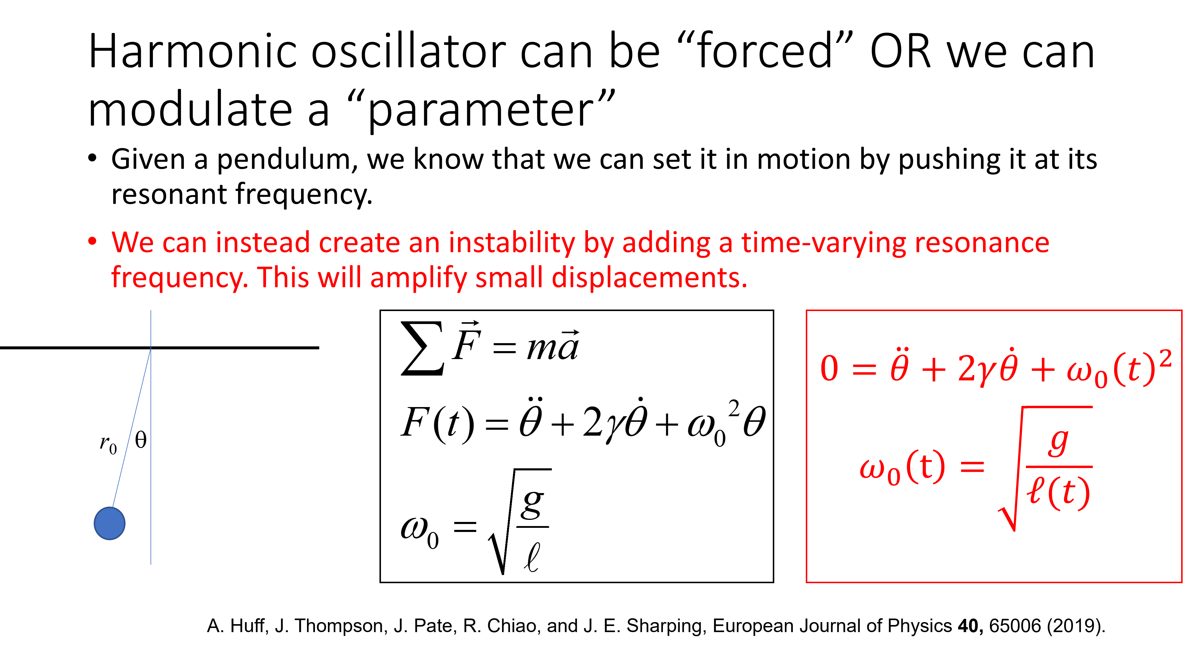

Parametric devices

Consider the superconducting radio-frequency (SRF) microwave cavity shown above. If the pump cavity is excited by microwave radiation resonant with the high-Q TE011 mode of the cylindrical cavity, energy efficiently builds up and radiation pressure acting on the movable membrane (shown in red) gives rise to periodic motion at twice the frequency of the pump. The dynamical Casimir effect (DCE) arises from the resulting time-varying boundary condition in the Signal/Idler cavity on the right-hand side of the membrane. The oscillating boundary, which acts like a moving piston, can do work upon vacuum fluctuations in the empty cavity such that the motion of the mirror can amplify these fluctuations into detectable radiation in the process of parametric amplification. Frequency-degenerate “Signal” and “Idler” fields (shown in yellow and having a frequency equal to that of the pump) build up within the high-Q cavity on the right-hand side. We calculate a pump power threshold for oscillation of ~100 uW, and we expect to observe laser-like generation of coherent states of radiation well above this threshold.

Applications of nonlinear fiber optics
The objectives of this program are to develop practical fiber optical parametric oscillator (FOPO) systems and to demonstrate their utility for stimulated Raman scattering (SRS) microscopy. Stimulated Raman scattering microscopy probes the Raman vibrational modes of molecules within an object and uses the signal to produce chemical specific images without the need to introduce fluorescent markers. When two optical fields pass through a material energy can shift between them. The transfer is efficient when the frequency difference between the two fields equals a Raman active mode frequency. The combination of spectroscopic resolution and nonlinear optics implies that there is a time-bandwidth tradeoff which, in this case, means that transform limited pulses of a few picoseconds in duration are optimal. Pulsed excitation, modulation and lock-in detection, and laser scanning permit the generation of high fidelity images at video acquisition rates.

Bringing SRS capability to a larger community. Currently, SRS microscopy is performed only in research laboratories whose specialty is developing and using novel imaging techniques. The high cost of SRS system components precludes its use in a wider range of scientific research laboratories. Moreover, complexity and size preclude applications to clinical research settings. The cost of commercialized versions of this technology will be determined by the marketplace, but fiber-based systems generally cost about one-third to one-half as much as a traditional bulk laser system. Several industrial partners have expressed an interest in our proposed system and we anticipate that commercial development will follow rapidly on meeting the performance benchmarks set forth within this proposal.
Fiber optical parametric oscillators (FOPOs). State of the art pulsed FOPOs utilize the nonlinear optical (Kerr) response of glass to generate wavelength-tunable, synchronous pulse trains in a manner similar to standard optical parametric oscillators. They are typically pumped by mode-locked lasers to obtain the peak powers necessary for operation. In order to build a truly portable system capable of generating suitable laser pulses we externally modulate the amplitude of a CW laser to produce pulses on the order of 20 ps in duration. After amplifying these pulses in two stages including a preamplifier and a high-power fiber amplifier they are directly coupled into a FOPO cavity using standard aspheric lenses. Cavity feedback is provided by the Fresnel-fiber reflection on one end of the Fabry-Perot cavity and a dichroic mirror output coupler on the other end. This should provide roughly 2%-4% feedback which is sufficient to provide stable operation at fairly high power levels. The wavelength is tuned by tuning the pump wavelength and by translating the output coupler to compensate for cavity dispersion. Because the system can tolerate high loss within the cavity one can use photonic crystal fibers (PCFs) that have been spliced to standard fibers, and further fiber integration is possible. We have demonstrated similar systems capable of delivering few-ps pulses with greater than 80 mW of average power. The PCF for our proposed system is slightly less than 2-m in length, and several commercially available PCFs can be used. In particular, the NL-1060 fiber from Crystal-Fibre is a good candidate.
FOPOs for CARS microscopy. These novel ultrafast light sources will significantly reduce the cost of equipment used to acquire chemical specific images. Here we observe the ability to chemically resolve lipid vesicles within a cell.

Optical trapping
By using a fiber-optical cell stretching apparatus, can one controllably, reversibly, and in real time, trigger the exchange of material across the phospholipid membrane? Can we use such a scheme to tailor the contents of giant vesicles for micro-reaction or drug delivery purposes? Does such a scheme provide a unique platform with which to explore the toxic effects of nanoparticles on living cells? In this proposal, we seek answers to these questions by optically stretching giant unilamellar vesicles (GUVs) whose membranes are doped with fluorescent or other functional proteins.

In one fundamental study we describe stable and unstable trapping regions for spherical dielectric particles in a dual-beam fiber optical trap. We find that, for large fiber separations, greater than twice the Rayleigh range of the trapping beams, one can stably trap particles whose sizes are comparable to or smaller than the beam size. If the particle size increases, unstable equilibrium causes particles to move away from the center of the trap towards the tips of the fibers. We also find that relatively large particles (with respect to the beam size) can be trapped when the fiber separation is less than twice the Rayleigh range of the Gaussian trapping beams. Most importantly, low refractive index contrast particles in a trap with small separation experience longitudinal trapping efficiencies and stretching forces which are one order of magnitude larger than in the conventional configuration.

GUVs are useful as a model system. The lipid bilayer is a semi-permeable barrier separating the interior of a biological cell from its exterior environment and compartmentalizing organelles from the cytoplasm. Cell stretching has been previously achieved in a dual fiber trap but we are the first to explore GUVs. The use of GUVs in our research program is essential because it allows us to isolate the mechanical contributions of the membrane from the contributions of other cellular components. Conformational changes in the lipid membrane itself play a surprisingly active role in cellular functions, and understanding fundamental lipid membrane mechanical properties will lead to better understanding the role of complex protein-membrane mediated interactions in proper cellular function. Beginning with a simple GUV we can then add complexity in a controlled fashion by introducing different lipid or protein components in the bilayer. The outcome is a versatile experimental system that can be used to carefully probe cell membrane mechanics.


